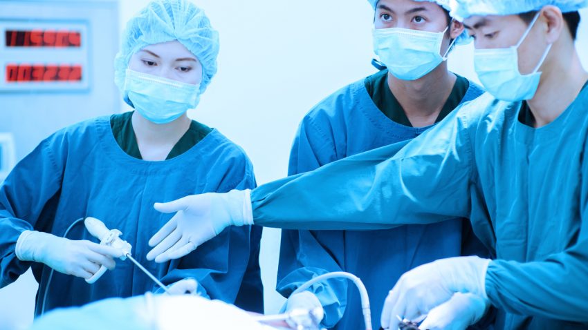

What Are Trocars? The Different Types & Their Uses in Surgery
Everything Health Professionals and Their Patients Need to Know About Laparoscopic Surgery
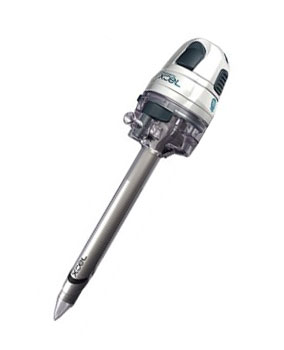
Trocars are a specialized medical device that can be used in a variety of scenarios. Most frequently, they are used during laparoscopic (keyhole) surgery, which is a procedure designed to cause minimal invasion to a patient requiring abdominal surgery. However, they may also be used to treat patients with hydrothorax or ascites and are utilized during the embalming process to remove gas and fluids from the body.
There are three main sections to a trocar:
- The cannula. This is the shaft that is inserted into the patient to give access to the abdominal cavity.
- The seal. This is found at the top of the cannula and ensures that no air escapes from the abdominal cavity while allowing any necessary equipment to be passed through. If air pressure is not properly maintained during laparoscopy, this can impede the surgeon’s view of the area being operated on.
- The obturator. This is the mechanism that enables the cannula to make the initial penetration into the abdomen.
In this article, we cover everything healthcare professionals and patients need to know about trocars, including their invention and development, how they are used in laparoscopic surgery, the conditions laparoscopic surgery is used to treat, and the risks and benefits of laparoscopy as well as how to prepare for laparoscopy and what to expect afterwards.
A Brief History of Trocars
Reginald S. Southey (1835-99) was the son of a doctor and followed in his father’s footsteps to enter medical practice. Based in London, England, he studied at Oxford and then St. Bartholomew’s Hospital, as well as in Europe, before subsequently working at London’s Hospital for Diseases of the Chest, where he developed a particular clinical interest in the kidney.
At the time, there were many medical and surgical instrument makers based in all the major cities who would make specific tools for individual medical professionals on request. Typically, a doctor would come up with an idea for an item that would solve a particular medical problem and put together a prototype using homemade equipment or modifying an existing piece of kit. They would then go to an instrument maker who could create a better prototype. If the new tool proved useful – or in some cases, if the inventor was influential – copies could then be readily made available through the instrument maker.
Dr Southey had worked with many patients suffering from anasarca (generalized swelling caused by an excess of fluid building up in subcutaneous connective tissue). At the time, the treatment for this was to wrap the affected area tightly with cloth, an uncomfortable and awkward method that severely hampered the patient’s ability to move around.
It is believed that Dr Southey probably observed fluid leaking from blisters on the affected area, which may have been the inspiration for his invention, Southey’s Tubes. He inserted a rigid tube subcutaneously, which enabled the excess fluid to drain away through a piece of rubber tubing into a bowl, providing relief to the patient.
Over time, he improved on his initial design, adding the trocar along with a skinny cannula and later making more holes along the tube in addition to the open end. Rubber tubing was frequently replaced with silver for repeated use.
The design was further amended to make it easy for doctors to carry it around when they made house calls. In these portable versions, the trocar handle had a screw-on top with holes drilled inside so that tubes could be placed in it along with the trocar, making it very simple to carry in a waistcoat pocket, so it was to hand whenever needed.
In the early twentieth century, William Green & Sons published their Encyclopedia and Dictionary of Medicine and Surgery. The eleven volume text offers invaluable insight into medical practices between the turn of the century and World War I, including the use of trocars.
One example given is their use in paracentesis. The text describes how a large trocar and cannula can be used to drain fluid quickly or, if a more gradual release is preferred, a smaller trocar, cannula and tubes should be chosen. It states that using finer equipment reduces the risk of fainting and is less distressing for the patient. There is a detailed description of how to use the equipment, stating that once the trocar has been inserted, the needle should be withdrawn and the cannula fixed in place with bandages, where it may remain for up to 24 hours. The book recommends using an abdominal bandage, gradually tightening it as fluid drains away, and then when the cannula is finally removed, a dressing should be applied to the wound. The final conclusion is that Southey’s tubes are superior to an aspirator when treating ascites (an abnormal build-up of fluid in the abdominal cavity).
Southey, who suffered from tuberculosis, had a personal interest in the process of draining pleural effusions. Fortunately, his tubes proved to be useful in this process as well. Although it is unclear in which order the various applications of the tubes came about, it is documented that Dr Southey reported on using his tubes to drain chests at a meeting of the Clinical Society of London in 1879 and his treatment enabled him to return to practice following his illness.
By the time Dr Southey passed away, his invention was stocked by the majority of medical suppliers and its use became commonplace in medicine. However, as diuretics improved as an alternative to manual drainage, later followed by tablets that were just as effective, Southey’s tubes were gradually phased out, leaving us with the modern day trocar.
A Background to Laparoscopic Surgery
Although many people think of laparoscopic (keyhole) surgery as a modern innovation, the first recorded use of the technique was by Albukasim, an Arabian physician who was born in the tenth century. However, it wasn’t until 1805 that this technique was explored further, when Phillip Bozzini utilized an illuminated light chamber, mirrors and a tube to view the urinary bladder, stones and neoplasms.
However, laparoscopic surgery continued to be overlooked in favor of traditional techniques until the early twentieth century, when German Georg Kellig used laparoscopes to examine the peritoneal cavities of dogs. Although Kellig claimed to have carried out the procedure on humans, the first documented instance of laparoscopy on human patients was by Hans Christian Jakobeus, a Swedish medic. He observed numerous pathologies using the technique and was able to detail a range of conditions, including cirrhosis of the liver, tuberculosis peritonitis and metastatic cancer.
One of the reasons why it took so long for laparoscopy to become widespread was the need for a source of light that didn’t burn the patient, as well as lenses that would enable the surgeon to have an unimpeded view. It was not until 1929 when a new lens system was developed that laparoscopy began to grow in popularity. This coincided with the development of the dual trocar method, allowing surgeons to view the abdominal cavity and easily get surgical instruments to where they were needed, marking a massive leap in laparoscopic techniques.
As laparoscopic surgery grew in popularity, it was initially used by gynecologists for basic procedures. It wasn’t until the early 1990s that general surgeons saw its value in gallbladder operations. As laparoscopic gallbladder surgery increased in popularity, the technique spread to other areas, such as colon surgery.
Laparoscopic technology and methods are continually improving and evolving, with modern innovations making this technique even more popular.
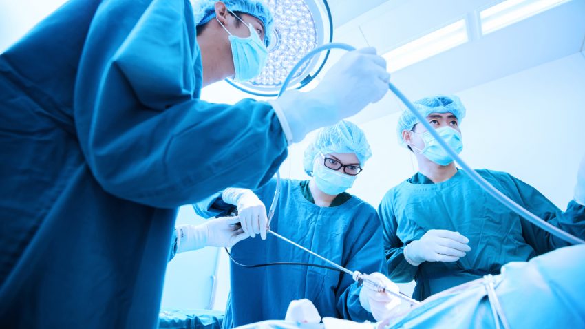
Laparoscopic surgery
The Benefits of Laparoscopic Surgery
These days, laparoscopic surgery is a common surgical approach, utilizing small incisions combined with specially adapted instruments to examine the organs located in the abdomen. Diagnostic laparoscopy minimizes scarring and distress to the patient. Recovery from laparoscopic surgery tends to be faster and there is less pain than with traditional surgical methods, and many procedures such as hernia repairs, gastric bypass, and organ removal, are all typically carried out laparoscopically. As well as offering many benefits to the patient, laparoscopy is also useful for doctors, who are able to see directly inside the body in real time without the need to perform open surgery. In addition, they can collect biopsy samples using this method when needed.
When is Laparoscopy Carried Out?
Laparoscopy is most frequently used to pinpoint and diagnose the cause of unexplained abdominal or pelvic pain. It is usually performed after other, non-invasive techniques, such as ultrasound, or CT scans, fail to provide adequate information to give a diagnosis.
A patient may be advised to have a laparoscopy to examine:
- The appendix
- The gallbladder
- The liver
- The pancreas
- The small and/or large bowel
- The spleen
- The stomach
- The pelvic area
- The reproductive organs
Diagnostic laparoscopy will allow the doctor to look for any unusual masses or tumors; fluid build-up in the abdominal cavity; liver disease; and how far a cancer has progressed. In addition, it can also demonstrate how effective a particular treatment has been.
The Risks Associated With Laparoscopy
One of the reasons why laparoscopic surgery is so popular is because it has relatively few risks compared to more traditional techniques. However, as with any surgery, there is still the chance of complications, which a [patient will need to understand before consenting to the procedure.
The most common side effects are bleeding and infection, but these are very rare. Patients should be made fully aware of any possible signs of infection. If someone has undergone a laparoscopic procedure and experiences any stomach pain that intensifies over time rather than subsiding, chills or fever, any redness, bleeding or oozing at the incision site, nausea or vomiting, coughing, difficulties breathing, problems urinating or light headedness, they should be advised to seek medical attention immediately.
There is also a tiny risk of damage to the abdominal organs during laparoscopy, which could cause blood or fluids to leak into the abdominal cavity. In the unlikely event of this occurring, further surgery is required to repair the damage. Finally, as with other surgical procedures, there are also risks of complication from the anesthesia or the chance of a blood clot, as well as the possibility of inflammation of the abdominal wall.
Laparoscopic surgery has been vigorously studied in a wide range of clinical trials across the globe. Overall, the conclusion has been that outcomes from minimally invasive surgery are comparable to traditional open surgery. There are occasions when a surgeon will discover conditions during the surgery that would make continuing with a minimally invasive approach unwise. When this occurs, the incision is enlarged so that the operation can be completed using traditional surgical techniques. This is known as a ‘conversion’ to traditional surgery and is not considered to be a complication of surgery so much as a demonstration of good judgment on the part of the surgeon.
If a patient is concerned about the possibility of conversion, they should discuss with their surgeon the risks associated with the procedure they are having in conjunction with their personal medical history.
What Happens During Laparoscopy?
Due to the fact that laparoscopy is a minor procedure with minimal risk, it is usually carried out as an outpatient appointment, meaning there is no need for an overnight stay following surgery. It can be done at a hospital or outpatient surgical center.
Laparoscopy is usually performed under general anesthesia, so the patient will be asleep throughout the operation and not feel any pain. However, in certain circumstances, local anesthesia is used, numbing the site of the operation so that although the patient is awake and aware, the procedure is still painless.
Once the patient has been anesthetized, a small incision is made just below their belly button and a trocar is thread through the hole. Carbon dioxide gas is fed through the trocar to inflate the abdominal cavity so the doctor has a clear view of all the internal organs. Once this is done, a laparoscope is placed through the cut with a camera attached so that the doctor can view the organs on a screen during the procedure.
Depending on requirements, up to four incisions will be made to allow for other surgical instruments, e.g. the tools required for a biopsy, which is when a small tissue sample is removed for testing. Laparoscopic surgical stapling tools allowing surgeons to divide and reconnect tissue have been developed, as have energy tools for cutting and cauterizing tissues and blood vessels. For some operations, a slightly larger incision may be necessary (around 2-4 inches long) so that tissue can be taken from the abdomen.
After the surgery is finished, all the instruments will be removed and the incisions stitched or taped closed. The patient may be bandaged up to protect the wounds.
Although laparoscopy is usually carried out on an outpatient basis, the patient will be required to remain in the healthcare unit for a few hours for monitoring. Their vital signs, such as heart rate and blood pressure, will be checked regularly and they will also be watched for signs of bleeding or adverse reactions to the anesthesia. Depending on their personal circumstances, they may need to stay overnight to make sure that they are well enough to go home. Alternatively, if they are discharged on the same day as the surgery, they will need to make arrangements for someone to take them home, since it is unsafe to drive in the hours following general anesthesia.
It is normal to feel slight pain and/or throbbing at the site of the surgery for a few days. Painkillers may be prescribed or the physician may advise on over-the-counter options. Shoulder pain is also a normal after effect, which is an effect of the carbon dioxide gas irritating the diaphragm which shares nerves with the shoulder, as is mild bloating. These should all go away after a few days.
The patient should be able to resume their normal daily activities within a week of the procedure. They will also need to visit your doctor two weeks after your laparoscopy for a follow up appointment.
The Various Types of Laparoscopic Surgery
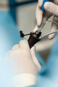 ‘Laparoscopic-assisted surgery’ is the term for a procedure that is carried out using predominantly laparoscopic techniques before being completed via a small abdominal incision. Many so-called laparoscopic surgeries are in fact technically laparoscopic-assisted, since some of the operation is completed through this specimen-removal incision.
‘Hand-assisted laparoscopic surgery’ involves making a small incision of around 2-3 inches into which a device is placed. This device enables the surgeon to place their hand inside the abdomen for assistance during the operation. This is done in conjunction with a laparoscope to watch proceedings on monitors, so that the surgeon can see what is happening in real time, and utilizes the same medical instruments as in traditional laparoscopic procedures.
This approach is popular for those instances when it is considered useful for the surgeon to be able to use their hand during the operation and collected specimens are removed through the same device that enables the surgeon to place their hand inside the abdomen. However, it has the disadvantage that in order for the hand to be able to pass into the abdomen, a larger incision is required than with traditional laparoscopic surgery. Nevertheless, studies report that hand-assisted laparoscopic surgery offers the same recovery outcome as pure laparoscopies.
‘Single incision surgery,’ otherwise known as ‘single site surgery’ or ‘laparoendoscopic single-site surgery’ (LESS) involves passing both the laparoscopic camera and any required medical instruments through a single incision approximately 2 inches long to remove a sample. The obvious advantage to this method is reduced scarring, since there is no need for extra trocar incisions. It has also been reported that it offers a faster recovery time with less need for postoperative analgesics.
However, many surgeons find this technique more challenging than other types of laparoscopic surgery since the instruments are by necessity placed very close to each other. In addition, it is only possible to get a two-dimensional view of the operating area and it is difficult for the surgeon to get into a good position with only one operating site. Finally, there additional training is required to master this technique, especially for suturing.
The technical challenges involved in LESS have meant that although it was first pioneered in the 1970s for tubal ligations, it failed to gain in popularity until more recent years. Advances in flexible optical and coagulation devices have made more complex procedures easier to carry out using LESS, such as hysterectomies and pelvic/aortic lymphadenectomies. Over the past decade, studies have also been carried out to show the usefulness of this approach in treating benign and malignant uterine disease.
‘Robotic surgery,’ sometimes called ‘robotic-assisted surgery’ is a relatively recent innovation and is frequently used in colon and rectal surgeries. It uses a similar technique to traditional laparoscopic surgery, where trocars are used to feed instruments into the abdomen, but rather than operating directly, the surgeon sits at a console with specialist computing equipment where they manipulate the instruments remotely while watching proceedings on a 3D monitor.
Since the robot can only work on a relatively small part of the abdomen at any given time and is hard to reposition, it is usually used for just a part of the operation, with the remainder of the procedure carried out using traditional laparoscopic techniques. However, robotic surgery is becoming increasingly popular, particularly in rectal operations, since robotic instruments have been designed to work well in the pelvic area where it is difficult to carry out diagnostic laparoscopy.
As a new technique, fewer studies have been carried out comparing the outcomes of robotic surgery to other types of minimally invasive surgery or traditional methods. It has been hypothesized that the benefits include greater visibility and an improved ability to carry out specific procedures on difficult to access parts of the body. However, the cost of the robot is currently very high, so not all health care facilities have the budget to offer robotic surgery and extra training is required for surgeons to be able to use the equipment with confidence.
‘Laser laparoscopic surgery’ is a term that many patients have heard of, but this refers to earlier approaches to laparoscopic gallbladder surgery. Lasers are no longer used in minimally invasive surgery, although this may change as technology develops.
The Different Types of Trocars
Modern trocars or abdominal access systems are available in a wide range of sizes from 3mm to 12mm and above and can be divided into two main categories:
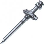
Bladed cutting trocar
- Cutting trocars, which have a sharp metal or plastic blade that can cut through the various tissue layers as pressure is applied. This type allows for easy insertion into the abdominal category.
- Dilating trocars, which have a blunt tip that separates and dilates tissue under pressure. These dilating trocars are noncutting and are a recent innovation aimed at removing the blade necessary for cutting systems, minimizing the risk of cutting internal organs.
However, there are a wide range of trocars available within those two categories designed to offer specialist features depending on need, for example, working ports, camera ports, and static or retraction ports. Choosing the right trocar depends on the nature of the procedure being carried out. For straightforward surgeries, e.g. diagnostic laparoscopies, a 5mm optic trocar is usually more than adequate to give enough light and control. More complicated procedures necessitate brighter light and a clearer picture, so a 10mm optic trocar is likely to be a better choice.
Standard procedures require two working trocars on either side of the lower abdomen and 5mm ports are most often used to allow access for secondary instruments. If a procedure is without complications, a smaller size can be utilized, while larger trocars of 12mm or above are used for those circumstances when a morcellator is required or large tumors are removed.
Disposable trocars are increasingly popular, offering the benefit of a guaranteed sharp tip requiring less pressure to insert. However, this is offset by the increase in cost, as well as the environmental impact.
In contrast, reusable trocars come with two different types of tip – conical and pyramidal. Pyramidal is the sharper of the two and therefore more popular. Reusable trocars are more economic than reusable, but require cleaning and sterilizing as well as regular sharpening and maintenance.
How Patients Can Prepare for Laparoscopic Surgery
If you have been advised that you need laparoscopy, it can be a frightening prospect, especially if you have never had surgery of any type before. It is worth preparing yourself in advance with as much information as possible so that you are aware of what is involved and the possible aftermath.
Familiarize yourself with the potential risks of your recommended laparoscopic surgery and ask your surgeon about the alternatives so that you can consider the risks vs the benefits of all your options. Make sure you fully read all pre- and post-operative materials and speak to your physician about anything you find unclear or confusing so you have a full understanding of what to expect. Pay particular attention to the signs of an undetected trocar injury so you know what’s normal and what’s not so that you can seek help immediately if you notice any dangerous symptoms developing.
 If you are nervous about the surgery, take time out to analyse what is making you scared. Is it the risk of complications? The anesthesia? The pain? The results of any sample testing? Once you know what exactly you’re afraid of, you can work to minimize the problem. If you have concerns about your pain tolerance levels, speak to your doctor about pain management before the procedure so they can make sure you have everything you need to be comfortable.[1] If you are worried about the anesthetic, arrange to talk to your anesthesiologist and doctor before the operation so they can answer any questions and allay your fears. You may also find it helpful to listen to relaxation tapes to prepare you for surgery.
Everyone’s experience of laparoscopic surgery will be different depending on your expectations, the nature of the surgery, your health care facility and staff, how you react to pain, etc. In addition, although doctors can advise on your likely recovery time, this is only an estimate, and while many people are fully recovered after a few days’ rest, some people can need several weeks to feel that they are back to their old self again.
Your surgeon may have ordered a bowel prep for the night before. This will vary according to the individual surgeon’s recommendations, but as a general rule, you will be put on a liquid diet and have to undergo various preparations to evacuate your bowels. Although this is not a nice process to go through, it is essential if you are having any bowel work. Check with your doctor about which medications and vitamins are safe to take prior to surgery.
Although laparoscopy is generally an outpatient procedure, complications may necessitate an overnight stay. If you require a bowel resection, you might even need to stay for a few days, so be prepared to spend the night, just in case. Pack loose clothing to wear after the procedure, ideally something without a waistband if possible. You may also want to take with you some socks and house slippers for comfort, as well as some other night things, just in case.
Make sure that you have arranged for someone to drive you home once you have been discharged. If possible, find someone who is supportive and can help you get out of the car and into your home. If they are able to stay overnight to make sure you’re okay, even better. You may find that you are feeling disoriented and struggle to move around after the procedure, so having someone to stay can be invaluable. Expect to feel under the weather for the next few days, so arrange for someone to be on call to help you, even if they can’t physically stay in your home. You may like to ask them to manage your medications for you for a few days to make sure you take them when you need. You are also likely to need someone to prepare your meals for you until you are feeling better.
Recuperation Following Laparoscopy
You may be advised to avoid driving for the first two weeks after your procedure. You are also likely to told to avoid intercourse, tub bathing, douching, and swimming. Be kind to yourself for the first few days and don’t push yourself too hard. You are likely to be tired and need plenty of rest and naps. However, don’t take this as an excuse to stay in bed. You will recover faster if you get up and move around as much as you feel capable of.
You will probably discover that loose clothing is the most sensible choice for a few weeks after your surgery. Your abdomen is likely to be a little swollen, with the incision sites tender to the touch. Avoid clothing that is tight around your middle and if you don’t have anything suitable to wear, it is a good idea to purchase a few options before your surgery for your comfort.
Many people find themselves feeling unusually emotional after their surgery, a feeling that can last for a few weeks. It is normal to find yourself becoming anxious or agitated, crying without reason or even having nightmares. All of this will pass in time, but if you have any concerns, speak to your physician for reassurance.
You may feel numb where your incisions are. This is caused by the nerves being cut. This will gradually pass as the nerves heal and is a normal reaction. However, you should be aware of the possible signs of infection detailed earlier in this article. If you experience any of them, seek medical advice immediately. Women who have had a laparoscopy may find that their first menstruation after surgery is heavier than usual and more painful. This is perfectly natural and nothing to be concerned about. It takes longer to heal internally than externally, so your next few periods may be more painful than you are used to. However, if you are worried about the level of pain you are experiencing, contact your doctor.
Many patients find that it can be helpful to speak with others who have been through the same procedure. Look for local or online support groups for help and advice both before and following your surgery.
Avoiding Complications From Laparoscopic Surgery
There is a great deal that medical professionals can do to minimize the risk of injury associated with laparoscopic procedures.
Surgeons should familiarize themselves completely with any new device before using for the first time, including how to utilize the device, indications and contraindications and any warnings or cautions. This information may be found in the manufacturer’s written materials, CDs and DVDs, but can also be obtained by speaking to representatives from the manufacturer and talking to and observing colleagues familiar with the device.
Surgeons should carry out laparoscopic procedures under the supervision of a more experienced laparoscopist until they are completely at ease with the surgery and confident of their ability to carry out the procedure unassisted.
When deciding whether a patient is a suitable candidate for laparoscopy, they should fully assess the individual and ensure they are at low risk of complication. Alternatives to blind trocar insertion, e.g. laparotomy or Hasson method, should be considered for these types of patients:
- Patients with an existing history of abdominal surgery
- Children
- Small, thin adults
- Patients whose lower abdomen skin cannot be appropriately stabilized to allow safe insertion of the trocar, e.g. women who have had multiple pregnancies or patients with atrophic abdominal musculature
Before surgery commences, check that there are appropriate injury intervention mechanisms and personnel in place and during surgery be alert for any indication of injury. Should an injury occur, take appropriate action immediately. Medical professionals should also take care to position themselves and their patients in an ergonomic position during the surgery to cut back on the effects of fatigue.
All patients should be made aware of the risks associated with the use of trocars, as well as the warning signs of serious complications so that patients can take action should they suffer any adverse effect. In the case of a negative side effect associated with trocars, these should be reported according to institutional protocol or through MedWatch, the FDA’s voluntary reporting program, either by visiting the website or by calling 1-800-FDA-1088.
Staying Up to Date With the Latest Advances in Laparoscopic Technology
Laparoscopic surgery offers a multitude of benefits to patients, including minimized scarring, and with the latest advances, these benefits will increasingly outweigh any potential risk. We’ve come a long way since Southey’s tubes and there are more exciting new innovations and techniques being developed all the time.
It is important to maintain appropriate stocks of trocars to ensure that you have the right sized piece of equipment to suit all eventualities. Laparoscopic technology is still constantly changing and improving, so medical facilities should ensure that they upgrade to the latest technology to ensure consistent high standards of patient care, while surgeons should take care to make sure that they understand how to use any new devices, attending further training if necessary.
Do you use disposable or reusable trocars or both in your healthcare facility? Why? Where do you see the future of laparoscopic surgery heading? Let us know your thoughts in the comments.
Sources:
[1] http://www.thehealthyhomeeconomist.com/laparoscopic-surgery-doctor-question/
Further reading:
[1] https://www.ncbi.nlm.nih.gov/pmc/articles/PMC4664217/
[2] https://www.fda.gov/MedicalDevices/Safety/AlertsandNotices/ucm197339.htm


3 thoughts on “What Are Trocars? The Different Types & Their Uses in Surgery”
Comments are closed.